应用枢椎关节面下螺钉固定治疗椎动脉变异的疗效评估
摘 要:目的 评估采用寰枢椎关节面下螺钉固定治疗椎动脉变异的可行性和有效性。方法 2013年1月至2021年1月上海交通大学医学院附属第九人民医院骨科采取枢椎关节面下螺钉行寰枢椎后路固定的椎动脉变异患者17例。术前通过CT二维重建在冠矢轴三个平面上确定螺钉轨迹,测量最狭窄处的直径。随后根据轨迹确定螺钉进针点。在矢状位测量螺钉轨迹长度和头倾角度。在轴位测量内倾角度,同时测量峡部长度作为峡部螺钉轨迹的长度。结果 关节面下螺钉轨迹处的最小直径为(5.5±0.9)mm,显著高于椎弓根最小直径(P<0.05)。进针点距离寰枢椎关节面的距离为(2.7±0.4)mm。距离椎管内壁距离为(3.4±0.3)mm。螺钉轨迹头倾角度为(7.4±2.5)°,内倾角度为(12.2±2.1)°。关节面下螺钉轨迹长度为(20.4±2.4)mm,峡部螺钉轨迹长度为(16.9±2.6)mm,两者间差异有统计学意义(P<0.05)。结论 当进行寰枢椎固定时,对于因椎动脉变异导致枢椎椎弓根狭窄而不适合接受椎弓根螺钉固定的患者。枢椎关节面下螺钉可作为替代方案。术前CT重建测量有助于安全有效地植入螺钉。
关键词:枢椎;椎动脉;螺钉;
Clinical application of atlantoaxial subarticular screw fixation for vertebral artery variation
Cheng Xiaofei Yang Erzhu Tian Haijun Sun Xiaojiang Zhao Changqing Zhao Jie
Shanghai Key Laboratory of Orthopaedic Implants, Department of Orthopaedic Surgery,Shanghai
Ninth People's Hospital, Shanghai Jiao Tong University School of Medicine
Abstract:
Objective To evaluate the feasibility and efficacy of atlantoaxial subarticular screw fixation for vertebral artery variation. Methods From January 2013 to January 2021, clinical data were obtained from 17 patients receiving atlantoaxial fixation using atlantoaxial subarticular screws in Department of Orthopaedic Surgery, Shanghai Ninth People's Hospital. Two-dimensional CT reconstruction was used to determine screw trajectory. The diameter of the narrowest place, the distance of entry point to atlantoaxial articular surface and spinal canal, the path length and angle of the screw trajectory and the length of the isthmus interarticularis were measured. Results The diameter of the subarticular trajectory was(5.5±0.9)mm, which was significantly higher than the diameter of the pedicles(P<0.05). The distance of entry point to atlantoaxial articular surface and spinal canal was(2.7±0.4)mm and(3.4±0.3)mm, respectively. The insertion angle of screw was(7.4±2.5)° cephalad to the axial plane and(12.2±2.1)° medial to the sagittal plane. The length of the screw trajectory and the isthmus was(20.4±2.4)mm and(16.9±2.6)mm, respectively, with a significant difference between them(P<0.05). Conclusion Atlantoaxial subarticular screw could be utilized as an alternative when pedicle screw is not suitable for axial fixation due to vertebral artery variation and narrowing of the pedicles. Preoperative CT reconstruction measurements are necessary for safe and effective screw insertion.
Keyword:
Axis; Vertebral artery; Screw;
枢椎的常见内固定方式包括椎弓根螺钉、峡部螺钉和椎板螺钉,其中椎弓根螺钉由于技术成熟,目前应用最为广泛。椎弓根螺钉通常从枢椎下关节突后方进钉,斜向头端通过关节突间峡部和椎弓根后直达椎体前缘皮质,其中椎弓根是这一骨性通道中最狭窄的部分,其外侧偏前有椎动脉穿过。椎动脉在枢椎内的走行复杂,且经常发生解剖变异如椎动脉高[1,2]。有研究显示,椎动脉高跨在人群中的发生比例在10%~31%[3]。椎动脉变异除了高跨外还存在内移的情况,内移和高跨的比例可分别达到25.5%和24%[4]。当存在这些解剖变异时,椎动脉占据枢椎内更多的骨性区域,使得椎弓根变狭窄,植入椎弓根螺钉有穿透骨壁损伤椎动脉的风险[5],因此置钉前需要通过影像学检查仔细评估椎动脉在枢椎内的走行和椎弓根的直径。遇到椎弓根狭窄的情况,可以选择植入更加安全的枢椎峡部螺钉或椎板螺钉[6,7]。但有文献报道,这两种技术的固定强度和最终融合率低于椎弓根螺钉[8,9],术后仍需辅助外固定。有学者研究测量了269例患者的枢椎CT数据,发现椎弓根小于4 mm者不适宜接受常规椎弓根螺钉固定,但在椎弓根的上部及寰枢椎关节面之间的骨质则有可能容纳螺钉植入[10]。此后,Patkar[11]介绍了一组在此区域进行置钉的病例,但并未提及这些病例是否存在椎动脉变异和椎弓根狭窄的情况,也没有进行具体的术前影像评估和设计。当需要进行枢椎固定时,应首先进行术前CT测量,发现枢椎椎动脉变异和椎弓根狭窄时,进一步评估改用寰枢椎关节面下螺钉技术进行固定的可行性。
资料与方法
一、资料
2013年1月至2021年1月上海交通大学医学院附属第九人民医院骨科采取枢椎关节面下螺钉行寰枢椎后路固定的椎动脉变异患者17例。手术指征包括:寰枢椎不稳5例,齿状突骨折5例,齿状突游离小骨4例,寰枢椎脱位3例。男10例,女7例,平均年龄43.6岁。术前患者全部接受颈椎正侧位、椎动脉CT(CT angiography, CTA)和颈椎MRI检查。
二、方法
1. 图像测量:
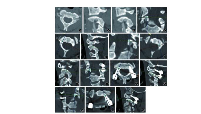
本组患者为因术前CTA发现存在椎动脉变异,不能植入椎弓根螺钉。为准确植入关节面下螺钉,对每一名患者的CT二维重建图像进行测量。首先在两侧的冠矢轴3个平面上确定螺钉中轴的最佳轨迹;随后测量骨质最狭窄处的直径,以此确定骨性通道是否可植入螺钉。根据轨迹确定螺钉进针点,在矢状位测量螺钉轨迹长度和头倾角度,在轴位测量内倾角度。同时测量双侧峡部长度,即矢状位下关节突中点到横突孔的距离,作为峡部螺钉轨迹的长度(图1 )。
2.手术:
术中根据术前测量结果,以关节间峡部背侧为进针点,寰枢椎侧块关节面下方约3 mm水平。用神经剥离子探及枢椎椎管内壁,取其外侧约3 mm进针。采用磨钻磨除进针点处少量皮质骨,开口后用开路器逐渐深入;方向为头端倾斜5°~10°,向内侧倾斜10°~15°。根据测深结果选择不同长度的3.5 mm直径螺钉植入(图1 )。寰椎侧块螺钉采用标准方法植入,随后用连接棒固定寰枢椎螺钉。最后行寰椎后弓及枢椎椎板去皮质,自体髂骨块植骨。术后第3~5天行颈椎正侧位及CT检查,评估复位情况和内植物位置。术后给予颈托固定2~3周。术后1年随访复查颈椎正侧位及CT检查观察内植物及植骨融合情况。

3.统计学处理:
采用SPSS 19.0软件进行数据分析。计量数据以x¯±s描述。两组间数据比较采用两样本t检验。P<0.05为差异有统计学意义。
结 果
术前CT显示该组患者全部存在枢椎椎弓根狭窄,直径为(3.2±0.5)mm, 不能接受椎弓根螺钉植入。在CT二维重建上测量关节面下螺钉轨迹的直径为(5.5±0.9)mm, 显著高于椎弓根直径(P<0.05),可以安全植入3.5 mm螺钉。设计进针点距离寰枢椎关节面的距离为(2.7±0.4)mm, 距离椎管内壁距离为(3.4±0.3)mm。螺钉轨迹头倾角度为(7.4±2.5)°。内倾角度为(12.2±2.1)°。关节面下螺钉轨迹长度为(20.4±2.4)mm。峡部螺钉轨迹长度为(16.9±2.6)mm, 两者间差异有统计学意义(P<0.05)。术后CT显示34枚枢椎螺钉均位于骨质内;其中7枚螺钉突破内侧骨皮质,横突孔处皮质完整,未损伤神经或椎动脉。患者随访1年以上,均实现骨性融合,未发现内植物失效情况。
讨 论
寰枢椎固定有助于实现节段的稳定和融合。常见的寰枢椎后路固定方法包括经关节突螺钉、寰椎侧块/椎弓根螺钉加枢椎峡部/椎弓根/椎板螺钉。经关节突螺钉可以提供良好的即刻稳定性。但单独应用这一技术时其抗屈曲应力能力有限,常需要与寰椎线缆或挂钩联合使用以降低屈曲时内固定失败风险,因此,这一技术应用于需要进行后弓切除减压的情况时需慎重。同时,这项技术不能进行术中的进一步复位,对术前复位要求较高。由于进针角度大,对于肥胖及胸椎后凸大的患者,理想的置钉存在一定困难。此外,螺钉通过枢椎时有损伤椎动脉的风险。
枢椎椎弓根螺钉最早被应用于枢椎Hangman骨折的后路手术治疗[13]。Goel等[14]首先采用寰椎侧块螺钉和枢椎椎弓根螺钉的钉板系统进行寰枢椎固定。随后Harms等[15]使用钉棒系统对其进行了改良。由于固定牢靠、适应范围广,同时可进行术中复位,目前这一技术已逐渐成为寰枢椎固定的主流术式。但临床研究的结果显示,枢椎椎弓根螺钉同样存在损伤椎动脉的潜在风险。Punyarat等[16]对术后CT分析发现植入的枢椎椎弓根螺钉中有23%(12枚/52枚)穿透骨皮质。Alosh 等[17]同样发现螺钉穿透皮质比例为23%,且椎动脉损伤的发生率为0.6%。一项荟萃分析纳入了2 975枚枢椎椎弓根螺钉的植入结果,发现椎动脉损伤发生率为0.3%[7]。
常规枢椎椎弓根螺钉走行通过下关节突、峡部和椎弓根。有学者通过影像学研究发现,椎弓根的宽度是制约椎弓根螺钉安全植入的主要因素,约有5%~9%的人群因为椎弓根过于狭窄,不适宜接受椎弓根螺钉固定[18,19]。而椎弓根狭窄的原因在于椎动脉的解剖变异,Yeom等[10]通过测量发现椎动脉变异比例达14.5%,相应的椎弓根狭窄(直径小于4 mm)比例达到9.5%。在椎动脉同时出现高跨和内移的情况下,椎弓根的平均直径为4.6 mm。为避免损伤椎动脉,植入椎弓根钉时需要增大外展角及头倾角。但即使这样的进针角度也很难植入常规椎弓根螺钉,因而推荐改用峡部螺钉或椎板螺钉[4]。
枢椎在解剖形态上与下颈椎完全不同,它的椎弓根因椎体形态差别和椎动脉走行变异存在很大的个体差异;其截面并非单纯的类圆形,普通轴位CT无法很好地评估椎弓根的形态和直径。对于接受上颈椎手术治疗的患者,术前常规进行椎动脉CTA检查。通过重建清楚地显示枢椎骨质形态和椎动脉走行,在设计螺钉的植入轨迹时才能有效避开椎动脉。
本组患者均存在椎动脉内移和/或高跨所致的椎弓根狭窄,椎弓根平均直径3.2 mm, 常规椎弓根螺钉轨迹会侵入椎动脉孔,具有血管损伤风险,不能接受常规椎弓根螺钉植入。因此,本研究选择枢椎关节面下螺钉作为替代方案。为准确植入螺钉,术前通过CT二维重建在冠矢轴三个平面上确定螺钉中轴的最佳轨迹,随后测量骨质最狭窄处的直径。经过测量。本组病例在椎动脉高跨和内移的情况下,椎动脉在枢椎内的走行为先内移再向前外侧高跨,因此椎动脉内侧的椎弓根比较狭窄,平均直径为3.2 mm。按常规方法植入椎弓根螺钉有风险,而在椎动脉沟的内上方的椎弓根骨质受椎动脉影响较小,平均直径为5.5 mm, 可以安全植入3.5 mm螺钉。
本组病例采取徒手方式,依据术前测量的结果进行置钉,术中使用C臂或O臂进行透视。术后CT显示螺钉位置均位于骨质内,未出现椎动脉损伤情况。关节面下螺钉的进针点高于常规椎弓根螺钉,位于侧块关节间峡部背侧,螺钉通过峡部和椎弓根内上方的骨质进入椎体内实现有效固定。由于进针点高并且螺钉走行远离椎动脉而朝向齿状突方向,可以避免螺钉侵入椎动脉孔。同时,螺钉通过了峡部和椎弓根,其固定形式和穿行轨迹与椎弓根螺钉相近。螺钉与椎弓根内侧及上方皮质骨形成直接接触,固定强度接近椎弓根螺钉。术前CT测量结果显示,本组关节面下螺钉的轨迹长度要显著大于峡部螺钉轨迹长度,提示其固定强度可能也高于峡部螺钉。
值得注意的是并非所有椎弓根狭窄病例都适合植入枢椎关节面下螺钉。对于椎动脉变异明显、术前CT测量显示预计螺钉轨迹直径小于4 mm的患者,仍无法安全植入关节面下螺钉,应改用其它固定方式[20,21,22]。
综上,当进行寰枢椎固定时,对于椎动脉变异伴枢椎椎弓根狭窄而不适合接受椎弓根螺钉固定时,寰枢椎关节面下螺钉可作为一个备选方案。术前通过CT重建测量有助于安全有效地植入螺钉。但本研究主要基于关节面下螺钉影像学测量结果,数据具有一定的局限性。此外,本研究并不能确定关节面下螺钉与椎弓根螺钉、峡部螺钉及椎板螺钉在力学性能上的差异,后续需要进一步进行尸体标本的测量和体外生物力学测试以明确这些问题。
参考文献
[1] Wakao N,Takeuchi M,Nishimura M,et al.Vertebral artery variations and osseous anomaly at the C1-2 level diagnosed by 3D CT angiography in normal subjects[J].Neuroradiology,2014,56(10):843- 849.DOI:10.1007/s00234-014-1399-y.
[2] Klepinowski T,Z yłka,Pala B,et al.Prevalence of high-riding vertebral arteries and narrow C2 pedicles among Central-European population:a computed tomography-based study[J].Neurosurg Rev,2021,44(6):3277-3282.DOI:10.1007/s10143- 021- 01493- 6.
[3] Yamazaki M,Okawa A,Furuya T,et al.Anomalous vertebral arteries in the extra-and intraosseous regions of the craniovertebral junction visualized by 3-dimensional computed tomographic angiography:analysis of 100 consecutive surgical cases and review of the literature[J].Spine,2012,37(22):E1389-E1397.DOI:10.1097/BRS.0b013e31826a0c9f.
[4] Lee,SH,Park DH,Kim SD,et al.Analysis of 3-dimensional course of the intra-axial vertebral artery for C2 pedicle screw trajectory:a computed tomographic study[J].Spine,2014,39(17):E1010-E1014.DOI:10.1097/BRS.0000000000000418.
[5] Elliott RE,Tanweer O,Boah A,et al.Comparison of screw malposition and vertebral artery injury of C2 pedicle and transarticular screws:meta-analysis and review of the literature[J].Clin Spine Surg,2014,27(6):305-315.DOI:10.1097/BSD.0b013e31825d5daa.
[6] Wright NM,Posterior C2 fixation using bilateral,crossing C2 laminar screws:case series and technical note[J].Clin Spine Surg,2004,17(2):158-162.DOI:10.1097/00024720-200404000- 00014 .
[7] Elliott RE,Tanweer O,Boah A,et al.Comparison of safety and stability of C-2 pars and pedicle screws for atlantoaxial fusion:meta-analysis and review of the literature:a systematic review[J].J Neurosurg Spine,2012,17(6):577-593.DOI:10.3171/2012.9.SPINE111021.
[8] Dmitriev AE,Lehman RA,Helgeson MD,et al.Acute and long-term stability of atlantoaxial fixation methods:a biomechanical comparison of pars,pedicle,and intralaminar fixation in an intact and odontoid fracture model[J].Spine,2009,34(4):365-370.DOI:10.1097/BRS.0b013e3181976aa9.
[9] Parker SL,McGirt MJ,Garcés-Ambrossi GL,et al.Translaminar versus pedicle screw fixation of C2:comparison of surgical morbidity and accuracy of 313 consecutive screws[J].Operat Neurosurg,2009,64 Suppl 5:343-349.DOI:10.1227/01.NEU.0000338955.36649.
[10] Yeom JS,Buchowski JM,Kim HJ,et al.Risk of vertebral artery injury:comparison between C1-C2 transarticular and C2 pedicle screws[J].Spine J,2013,13(7):775-785.DOI:10.1016/j.spinee.2013.04.005.
[11] Patkar SV.New entry point for C2 screw,in posterior C1-C2 fixation (Goel-Harm's technique) significantly reducing the possibility of vertebral artery injury[J].Neurolog Res,2016,38(2):93- 97.DOI:10.1080/01616412.2015.1105582.
[12] Chun DH,Kim KN,Yi S,et al.Biomechanical comparison of four different atlantoaxial posterior fixation constructs in adults:a finite element study[J].Spine,2018,43(15):E891-E897.DOI:10.1097/BRS.0000000000002584.
[13] Leconte P,Fracture T.Luxation des deux premie res verte bres cerviles:fracures du cou-de-pied ruchis cervical[J].Actualit Chirurg Orthopediq Raymon Poinc,1964,3:147-166.
[14] Goel A,Laheri V.Plate and screw fixation for atlanto-axial subluxation[J].Acta Neurochirurg,1994,129(1):47-53.DOI:10.1007/BF01400872.
[15] Harms J,Melcher RP.Posterior C1-C2 fusion with polyaxial screw and rod fixation[J].Spine,2001,26(22):2467-2471.DOI:10.1097/00007632-200111150- 00014.
[16] Punyarat P,Buchowski JM,Klawson BT,et al.Freehand technique for C2 pedicle and pars screw placement:is it safe?[J].Spine J,2018,18(7):1197-1203.DOI:10.1016/j.spinee.2017.11.010.
[17] Alosh H,Parker SL,McGirt MJ,et al.Preoperative radiographic factors and surgeon experience are associated with cortical breach of C2 pedicle screws[J].Clin Spine Surg,2010,23(1):9-14.DOI:10.1097/BSD.0b013e318194e746.
[18] Resnick DK,Lapsiwala S,Trost GR.Anatomic suitability of the C1-C2 complex for pedicle screw fixation[J].Spine,2002,27(14):1494-1498.DOI:10.1097/00007632-200207150- 00003.
[19] Yoshida M,Neo M,Fujibayashi S,et al.Comparison of the anatomical risk for vertebral artery injury associated with the C2-pedicle screw and atlantoaxial transarticular screw[J].Spine,2006,31(15):E513-E517.DOI:10.1097/01.brs.0000224516.29747.52.
[20] Lee DH,Park S,Cho JH,et al.The medial window technique as a salvage method to insert C2 pedicle screw in the case of a high-riding vertebral artery or narrow pedicle:a technical note and case series[J].Eur Spine J,2022,31(5):1251-1259.DOI:10.1007/s00586- 022- 07146- 6.
[21] Kothari MK,Dalvie SS,Gupta S,et al.The C2 pedicle width,pars length,and laminar thickness in concurrent ipsilateral ponticulus posticus and high-riding vertebral artery:a radiological computed tomography scan-based study[J].Asian Spine J,2019,13(2):290.DOI:10.31616/asj.2018.0057.
[22] Lu Y,Lee YP,Bhatia NN,et al.Biomechanical comparison of C1 lateral mass-C2 short pedicle screw-C3 lateral mass screw-rodconstruct versus Goel-Harms fixation for atlantoaxial instability[J].Spine,2019,44(7):E393-E399.


